 Akhil Ramnarain
Our planet is home to a wide array of biomes each booming with life of multitudes of colours. Yet the most vibrant of colours exist in the microscopic world. Bacterial pigments have vast applications namely their use in the medical, pharmaceutical , cosmetic and the food and beverage industry. Above all pigment producing microorganisms Pseudomonas aeruginosa is without a doubt my favourite. Allow me to convince you.
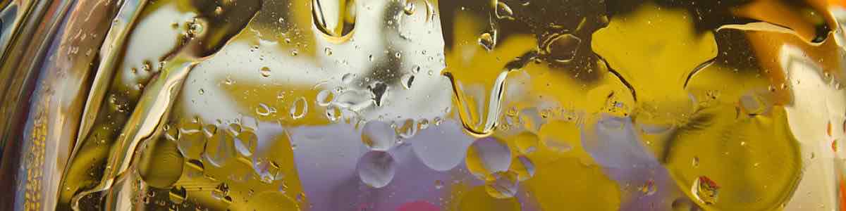
Just look at it, a true marvel of nature. Looks like something out of a sci-fi movie. Pseudomonas aeruginosa is made up of two pigments, pyocyanin (blue) and pyoverdine (green), which impart the blue-green characteristic color.
The attached magazine has an amazing article on page 14 on the use of microorganisms as a colour palette, who would have thought that a degree in microbiology could allow you to become an artist.
Sixty-three isolates belonging to the genus Pseudomonas were isolated from different environmental sources including; soil, water and clinical specimens. Twenty out of them were identified as Pseudomonas aeruginosa and individually screened for pyocyanin production. P. aeruginosa R1; isolated from rice-cultivated soil and P. aeruginosa U3 selected from clinical specimen (Urinary tract infection) were the highest pyocyanin producers; pyocyanin production reached 9.3 and 5.9 μg/ml, respectively on synthetic glucose supplemented nutrient medium (GSNB). The identification of both selected strains (P. aeruginosa R1 and P. aeruginosa U3) was confirmed by 16S rRNA, the similarity with other strains available in database was 97% (with P. aeruginosa FPVC 14) and 94% (with P. aeruginosa 13.A), respectively. P. aeruginosa R1 and P. aeruginosa U3 are accessed at gene bank with accession numbers KM924432 and KM603511, in the same order. Pyocyanin was extracted by standard methods, purified by column chromatography and characterized by UV-Vis absorption, mass spectrometry and nuclear magnetic resonance. The antimicrobial activity of purified pyocyanin against multi-drug resistant microbes was investigated; the efficiency of pyocyanin was more obvious in Gram +ve bacteria than Gram−ve bacteria and yeast. To reduce the cost of pyocyanin production, a new conventional medium based on cotton seed meal supplemented with peptone was designed. The pyocyanin production of both selected strains P. aeruginosa R1 and P. aeruginosa U3 using the new medium is increased by 30.1% and 17.2%, respectively in comparison with synthetic GSNB medium, while the cost of production process is reduced by 56.7%.
Author(s):
C. Cezard,
             N.  Farvacques,

                 P.  Sonnet.

    Laboratoire de Glycochimie, des Antimicrobiens et des Agroressources, CNRS FRE 3517, Universite de Picardie Jules Verne, UFR de Pharmacie, 1 Rue des Louvels, 80037 Amiens cedex 01, France., France
Pseudomonas aeruginosa is the most common pathogen that persists in the cystic fibrosis lungs. Bacteria such as P. aeruginosa secrete siderophores (iron-chelating molecules) and the host limits bacterial growth by producing neutrophil-gelatinase-associated lipocalin (NGAL) that specifically scavenges bacterial siderophores, therefore preventing bacteria from establishing infection. P. aeruginosa produces a major siderophore known as pyoverdine, found to be important for bacterial virulence and biofilm development. We report that pyoverdine did not bind to NGAL, as measured by tryptophan fluorescence quenching, while enterobactin bound to NGAL effectively causing a strong response. The experimental data indicate that pyoverdine evades NGAL recognition. We then employed a molecular modeling approach to simulate the binding of pyoverdine to human NGAL using NGAL’s published crystal structures. The docking of pyoverdine to NGAL predicted nine different docking positions; however, neither apo- nor ferric forms of pyoverdine docked into the ligand-binding site in the calyx of NGAL where siderophores are known to bind. The molecular modeling results offer structural support that pyoverdine does not bind to NGAL, confirming the results obtained in the tryptophan quenching assay. The data suggest that pyoverdine is a stealth siderophore that evades NGAL recognition allowing P. aeruginosa to establish chronic infections in CF lungs.
Pyocyanin is a biologically active phenazine produced by the human pathogen Pseudomonas aeruginosa. It is thought to endow P. aeruginosa with a competitive growth advantage in colonized tissue and is also thought to be a virulence factor in diseases such as cystic fibrosis and AIDS where patients are commonly infected by pathogenic Pseudomonads due to their immunocompromised state. Pyocyanin is also a chemically interesting compound due to its unusual oxidation-reduction activity. Phenazine-1-carboxylic acid, the precursor to the bioactive phenazines, is synthesized from chorismic acid by enzymes encoded in a seven-gene cistron in Pseudomonas aeruginosa and in other Pseudomonads. Phenzine-1-carboxylic acid is believed to be converted to pyocyanin by the sequential actions of the putative S-adenosylmethionine dependent N-methyltransferase PhzM and the putative flavin-dependent hydroxylase PhzS. Here we report the 1.8 Å crystal structure of PhzM solved by single anomalous dispersion. Unlike many methyltransferases, PhzM is a dimer in solution. The 36 kDa PhzM polypeptide folds into three domains. The C-terminal domain exhibits the α/β-hydrolase fold typical of small molecule methyltransferases. Two smaller N-terminal domains form much of the dimer interface. Structural alignments with known methyltransferases show that PhzM is most similar to the plant O-methyltransferases that are characterized by an unusual intertwined dimer interface. The structure of PhzM contains no ligands and the active site is open and solvent exposed when compared to structures of similar enzymes. In vitro experiments using purified PhzM alone demonstrate that it has little or no ability to methylate phenzine-1-carboxylic acid. However, when the putative hydroxylase PhzS is included, pyocyanin is readily produced. This observation suggests that a mechanism has evolved in P. aeruginosa that ensures efficient production of pyocyanin by preventing the formation and release of an unstable and potentially deleterious intermediate.
Antimicrobial Effect of Pyocyanin Extracted from Pseudomonas aeroginosa, Abdul-Hussein ZR and Atia SS
Biotechnology Laboratory, REDEC of Kajaani, University of Oulu, 88600 Sotkamo, Finland,1 and Laboratoire de Microbiologie et de Génétique, CNRS UPRES-A 7010, Université Louis-Pasteur, 67000 Strasbourg, France2


